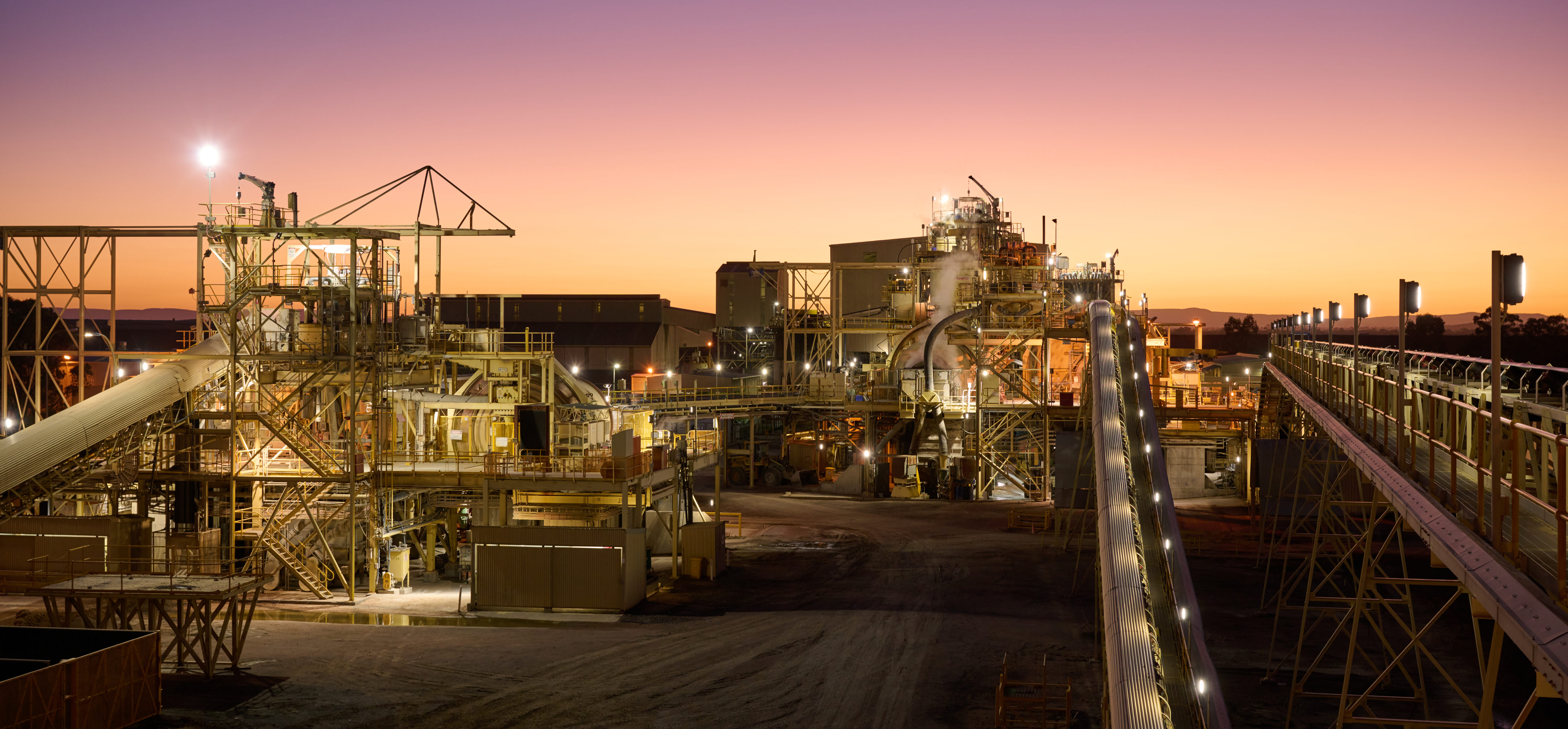
Copper could now be used for clearer MRIs

MRI. (Reference image by Mart Production, Pexels.)

Researchers at the Universities of Birmingham and St Andrews, as well as Diamond Light Source, discovered a novel copper protein binding site that has real potential for MRI contrast agents used to improve the visibility of internal body structures in scans.

The discovery overturns conventional medical wisdom that copper is unsuitable for use in MRIs and could help develop new imaging agents with fewer risks and side effects than those currently employed.

The experts found that the new structure displayed highly effective levels of relaxivity – the ability of a contrast agent to influence the relaxation times of protons – which helps create clearer and more informative images.

“Despite copper largely being disregarded for use in MRI contrast agents, our binding site was shown to display extremely promising contrast agent capabilities, with relaxivities equal and superior to the Gd(III) agents used routinely in clinical MRI,” Anna Peacock, co-author of the paper in Proceedings of the National Academy of Science that details the findings, said. “Our discovery showcases a powerful approach for accessing new tools or agents for imaging applications.”

Also for PET scans

According to Peacock and her co-authors, imaging agents based on copper could also be used in positron emission tomography (PET) scans, which produce detailed 3-dimensional images of the inside of the body.

Their study shows the use of an artificial coiled-coil to create the copper site within a protein scaffold has achieved function and performance not normally associated with copper.

“Metal sites that are not part of the repertoire of biology are vital in providing protein designers with an expanded toolbox of chemistries they can use to design new functional systems such as the promising imaging capabilities reported here,” Peacock said.

“This opens up applications beyond what biology is currently capable of and showcases some of the advantages of using simple miniature protein scaffolds as a means with which we can engineer new, and maybe currently unknown, metal-binding sites.”

The scientist explained that in MRI scanners, sections of the body are exposed to a strong magnetic field causing hydrogen nuclei of water in tissues to be polarized in the direction of the magnetic field. The magnitude of the spin polarization detected is used to form the MR image but decays with a characteristic time constant known as the T1 relaxation time.

Water protons in different tissues have different T1 values, which are the main sources of contrast in MR images. A contrast agent usually shortens but in some instances increases, the value of T1 of nearby water protons – altering contrast in the image and improving the visibility of internal body structures, with the most used compounds being gadolinium-based contrast agents.

Gadolinium (in the form of Gd3+) is often used as a contrast agent, but there are environmental and patient safety concerns making the exploration of new contrast agents an important and active research area.

Although more work needs to be done to secure the stability of this new copper protein site, the study authors believe their work is a promising first step toward designing new copper-based contrast agents for clinical MRI scanning.